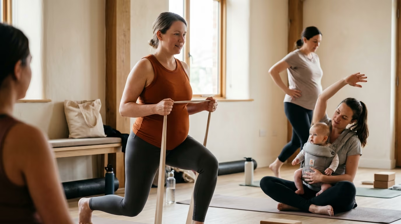

Navigating physical changes across trimesters and the fourth trimester requires adaptable strength routines that protect healing tissues and support maternal wellness.





Movement during pregnancy and the postpartum transition is not a performance test. It is also not a mandatory pause in physical development or a test of personal discipline. Instead, prenatal and postnatal movement is an adaptable process of matching physical activity to rapidly shifting physiological states. This guide examines how to maintain movement habits safely across every trimester and throughout postpartum recovery, prioritizing long-term health and tissue integrity over arbitrary fitness standards.
Physical activity during pregnancy and after childbirth supports cardiovascular function, metabolic health, and musculoskeletal resilience. Contemporary clinical evidence shows that structured strength and aerobic exercise reduce the risk of common gestational complications while improving physical comfort. By adopting a symptom-guided framework rather than a rigid calendar, individuals can sustain meaningful movement patterns while protecting their changing bodies.
Major international health organizations agree that regular physical activity is safe and beneficial for most pregnant and postpartum individuals. The World Health Organization recommends that pregnant and postpartum women without medical contraindications accumulate at least 150 minutes of moderate-intensity aerobic activity each week. This recommendation explicitly includes both cardiovascular conditioning and muscle-strengthening exercises. The Canadian Guideline for Physical Activity throughout Pregnancy mirrors this 150-minute recommendation, encouraging physical activity across a minimum of three days per week while highlighting the benefits of daily movement.
The American College of Obstetricians and Gynecologists, known as ACOG, affirms that physical activity during pregnancy presents minimal risks for individuals with uncomplicated pregnancies. ACOG emphasizes that physical activity helps manage blood glucose levels, improves psychological well-being, and maintains muscular strength. For the postpartum period, ACOG notes that physical activity can be resumed gradually as soon as it is medically safe. This resumption depends on delivery mode, surgical recovery, and personal comfort.
The scientific evidence supporting these guidelines is robust. Systematic reviews analyzing thousands of pregnancies demonstrate that regular exercise does not increase the risk of preterm birth, low birth weight, or miscarriage. In fact, accumulating research shows that resistance training and aerobic exercise reduce the odds of developing gestational hypertension by roughly 58 percent. Exercise also lowers the odds of developing gestational diabetes by approximately 38 percent. Furthermore, structured training reduces the risk of perinatal mood disorders and fetal macrosomia without causing maternal or fetal harm.
Despite these established targets, the 150-minute recommendation serves as a health benchmark rather than a strict pass or fail requirement. Physical capacity fluctuates based on sleep quality, morning sickness, musculoskeletal discomfort, and postpartum recovery demands. Some weeks may allow for full adherence to structured strength routines, while other weeks may only permit short walks and gentle mobility work. A person who accumulates three 10-minute bouts of movement in a day gains meaningful metabolic benefits. Moving when possible and resting when needed provides far greater long-term value than forcing adherence to an inflexible plan.
To train intelligently during pregnancy, one must understand the biological adjustments occurring within the body. Pregnancy involves profound hormonal, vascular, and mechanical adaptations designed to support fetal growth and prepare the body for birth. These changes directly influence how muscles produce force, how joints handle load, and how the cardiovascular system recovers from exertion.
The hormonal landscape changes dramatically from the first trimester onward. The hormone relaxin increases significantly, working alongside estrogen and progesterone to increase the laxity of ligaments throughout the body. This ligamentous softening is essential for allowing the pelvis to expand during delivery, but it temporarily reduces passive joint stability. As passive connective tissues become more compliant, the muscular system must work harder to stabilize the hips, pelvis, and lumbar spine. Strength training provides active neuromuscular support, compensating for this reduced ligamentous stiffness.
Cardiovascular physiology also undergoes massive shifts to nourish the developing placenta. Maternal blood volume increases by 40 to 50 percent, resting heart rate rises, and stroke volume expands. Because baseline cardiac output is elevated, maternal heart rate responds more rapidly to physical exertion. Resting blood pressure often drops during the second trimester due to decreased systemic vascular resistance, which can occasionally cause dizziness during rapid postural changes. The metabolic cost of movement also rises, meaning standard workloads demand higher oxygen consumption and energy expenditure.
Mechanical changes become pronounced as the uterus expands. The maternal center of mass shifts forward and upward, altering pelvic alignment and increasing the lordotic curve of the lumbar spine. This postural shift changes the mechanical leverage of the hip extensors and trunk musculature. The abdominal wall stretches to accommodate fetal growth, causing the rectus abdominis muscles to separate along the linea alba, a normal physiological adaptation termed diastasis recti. Concurrently, the pelvic floor muscles bear increased mechanical load from the weight of the uterus and changing intra-abdominal pressure dynamics.
Postpartum biology involves another distinct set of physiological processes. Following delivery, hormone levels drop precipitously, which can influence tissue recovery, joint laxity, and emotional stability. If a person is lactating, elevated prolactin and lower systemic estrogen levels can maintain joint laxity and alter vaginal tissue hydration for several months. Additionally, recovering from a vaginal delivery requires connective tissue and muscular healing in the pelvic floor, while recovering from a cesarean birth requires recovery from major abdominal surgery. Navigating these metabolic and physiological changes requires an understanding of evidence-led strength and fitness principles that prioritize gradual tissue loading.
A practical movement program requires clear safety parameters. Exercising during pregnancy is generally safe, but training must be adjusted or paused if specific health complications or symptoms arise. Clinicians categorize these considerations into absolute contraindications, relative contraindications, and immediate red-flag symptoms.
Absolute contraindications are medical conditions where strenuous physical activity presents clear clinical risks to maternal or fetal safety. According to the Canadian guidelines and ACOG, absolute contraindications include ruptured membranes, unexplained persistent vaginal bleeding, placenta previa after 28 weeks of gestation, preeclampsia, and an incompetent cervix. Other absolute contraindications include intrauterine growth restriction, higher-order multiple pregnancies such as triplets, uncontrolled type 1 diabetes, uncontrolled hypertension, and severe systemic cardiovascular or respiratory disorders. Individuals with these conditions must avoid structured exercise regimens, though basic daily activities may still be permitted under medical supervision.
Relative contraindications require individualized clinical discussion before engaging in moderate-to-vigorous physical activity. These conditions include a history of recurrent pregnancy loss, gestational hypertension, a history of spontaneous preterm birth, mild cardiovascular or respiratory disease, symptomatic anemia, malnutrition, and twin pregnancy after 28 weeks. A clinical consultation helps determine whether specific modifications, intensity caps, or specialized monitoring are necessary to keep movement safe and productive.
To simplify daily training decisions, exercisers can use a traffic-light decision model:
Movement is appropriate when an individual has medical clearance, feels physically energetic, and experiences no unusual discomfort. In this state, training can proceed with standard moderate-intensity resistance exercises, cardiovascular sessions, mobility work, and daily walking. The focus remains on consistent execution and progressive, manageable loading.
Modification is necessary when transient symptoms arise during or after exercise. These symptoms include joint discomfort, mild pelvic pressure, dizziness when changing positions, notable fatigue, or abdominal doming during exertion. Appropriate adjustments include reducing external load, shortening the range of motion, increasing rest intervals, changing body position, or choosing a more stable movement variation. For instance, shifting from a standing overhead press to a seated dumbbell press reduces balance demands and spinal shear forces.
Certain clinical signs require immediate cessation of exercise and prompt medical attention. ACOG defines these red-flag symptoms as:
These symptoms must never be ignored or trained through. Exercisers should stop their session immediately and contact their obstetric care provider or local emergency services based on symptom severity.
Intensity regulation during pregnancy requires flexible, subjective tools rather than rigid metrics. In non-pregnant training environments, lifters often rely on fixed heart-rate zones or percentages of a one-rep maximum. However, maternal resting heart rate, blood volume, and stroke volume fluctuate throughout gestation, making target heart-rate formulas unreliable.
The most dependable tool for monitoring cardiovascular and resistance intensity during pregnancy is the talk test. Endorsed by ACOG, the talk test relies on a simple physiological principle. If a person can carry on a comfortable conversation while moving, their exertion level is generally moderate and safe. If speaking requires gasping for breath between words, intensity has drifted into a vigorous or near-maximal zone that may require longer recovery.
Another effective tool is the Rating of Perceived Exertion, often measured on the Borg 6 to 20 scale or a simple 1 to 10 scale. A moderate intensity corresponds to a 12 to 14 on the Borg scale, which feels somewhat hard but completely sustainable. On a 1 to 10 scale, most prenatal strength work should sit between a 5 and a 7. This level of effort stimulates muscular adaptation and preserves joint health without creating extreme systemic fatigue. Prioritizing rest and tracking physical capacity aligns with comprehensive sleep and recovery strategies that protect maternal energy.
Recent research has challenged older assumptions regarding heavy resistance training during pregnancy. Historically, clinical guidelines discouraged lifting heavy weights due to theoretical concerns regarding reduced uterine blood flow or excessive pelvic pressure. However, a 2025 systematic review published in the British Journal of Sports Medicine found that prenatal resistance training did not increase adverse maternal or fetal outcomes. In fact, structured resistance exercise was associated with lower rates of gestational diabetes and preeclampsia.
Furthermore, studies examining experienced lifters who continued heavy strength training, including Olympic weightlifting and the Valsalva maneuver, observed normal perinatal outcomes. A 2025 clinical trial demonstrated that acute high-intensity resistance training with brief breath-holding was well tolerated by both mothers and fetuses in trained populations. However, these findings do not suggest that every pregnant individual should lift maximal loads or hold their breath under heavy strain. Rather, they show that strength training is fundamentally adaptable. For most individuals, keeping several repetitions in reserve and maintaining steady breathing ensures a safe and productive session.
Strength training during pregnancy does not require inventing an entirely new collection of exercises. Instead, it involves modifying fundamental human movement patterns to accommodate physical changes, protect joints, and manage intra-abdominal pressure. The primary movement patterns include the squat, the hinge, the push, the pull, the lunge, and the loaded carry.
The squat strengthens the quadriceps, gluteal muscles, and pelvic floor, supporting daily tasks like sitting down and picking up objects. As the abdomen expands and the center of mass shifts, a barbell back squat can place unnecessary strain on the lower back. Excellent prenatal modifications include:
Hinging exercises target the posterior chain, including the hamstrings, glutes, and spinal erectors. Maintaining posterior chain strength helps counterbalance the anterior pull of an expanding belly. Modifications include:
Upper-body pushing and pulling preserve shoulder health, support upright posture, and prepare the upper body for the physical demands of holding an infant. As pregnancy advances, lying flat on the back can cause the heavy uterus to compress the inferior vena cava. This compression reduces venous return to the heart, leading to dizziness, nausea, or lightheadedness, a condition termed supine hypotensive syndrome.
Unilateral leg training is exceptionally valuable for pelvic stability and balance, but it must be adapted if pelvic girdle pain or pubic symphysis dysfunction occurs. Widely split stances can create asymmetrical shear forces across the pelvis.
Traditional direct abdominal training like sit-ups and rotational twists can increase abdominal wall doming and excess intra-abdominal pressure. Instead, core training should emphasize anti-extension, anti-lateral flexion, and anti-rotation.
To help implement these adjustments throughout gestation, consider this trimester-by-trimester adaptation overview:
Returning to exercise after childbirth requires patience and structured progression. The common belief that individuals are entirely restricted for six weeks and then suddenly cleared for unrestricted exercise does not align with modern rehabilitation science. Tissue healing, hormonal recovery, sleep deprivation, and pelvic floor adaptations follow a continuum rather than an abrupt calendar date. ACOG notes that light physical activity can begin within days of an uncomplicated vaginal birth, while formal strength and high-impact routines should be rebuilt in stages.
A staged return model allows individuals to safely progress from basic functional recovery to demanding athletic endeavors:
The initial weeks following birth focus on circulation, tissue healing, and nervous system recovery. The primary goals are gentle movement, comfortable posture during infant feeding, and reconnecting with the deep core and pelvic floor.
Once basic daily tasks are comfortable and any perineal or cesarean incisions have begun healing, low-load resistance exercises can be introduced. ACOG explicitly recommends introducing exercises that strengthen the abdominal and back muscles to support maternal posture and daily infant care.
In this stage, external loads are progressively increased. Exercises progress from bodyweight movements to dumbbells, barbells, and cable machines.
High-impact activities such as running, jumping, sprinting, and maximal weightlifting place substantial mechanical demands on the pelvic floor and connective tissues. Clinical return-to-running guidelines recommend delaying high-impact running until at least 12 weeks postpartum, provided the individual is completely symptom-free.
Readiness for high-impact exercise should be evaluated using functional physical tests rather than arbitrary timelines. Before returning to running or jumping, an individual should be able to:
For additional structured strategies on physical progression, readers can review our collection of evidence-based movement resources.
Pelvic floor and abdominal wall health are central components of postpartum recovery. The pelvic floor is a sling of muscles and connective tissue that supports the bladder, uterus, and bowel, maintains continence, and manages intra-abdominal pressure. Pregnancy itself, regardless of delivery mode, places prolonged mechanical stress on these tissues. Childbirth can further stretch or tear muscular and neural structures.
Pelvic floor dysfunction is common, but it should never be dismissed as an unavoidable or permanent consequence of motherhood. Symptoms represent vital clinical data indicating that the body is struggling to manage current loads or pressure strategies.
Key pelvic-health symptoms that warrant evaluation by a pelvic-health physical therapist include:
A specialized pelvic-health physical therapist provides comprehensive internal and external evaluations. They assess pelvic floor muscle strength, endurance, resting tone, and coordination. Many people assume that pelvic symptoms always indicate a weak pelvic floor requiring Kegels. In reality, many individuals develop hypertonic, overactive pelvic floor muscles that cannot relax effectively. In such cases, performing additional Kegels can worsen symptoms. A physical therapist provides tailored cues, teaches breathing strategies, assists with cesarean or episiotomy scar mobilization, and crafts a personalized rehabilitation plan.
Misinformation regarding pregnancy and fitness can cause unnecessary fear, leading individuals to avoid beneficial movement. Addressing these common myths encourages informed, confident decision-making.
A persistent myth claims that lifting weights restricts fetal growth, causes miscarriage, or induces preterm labor. Modern clinical research shows that resistance training is safe and protective. Resistance exercise improves insulin sensitivity, reduces lower back pain, and helps maintain physical independence. Unless absolute medical contraindications exist, pregnant individuals can safely participate in appropriately scaled strength training throughout gestation.
Many people believe that receiving routine clinical clearance at the six-week postpartum visit means they can immediately resume intense workouts, heavy lifting, or running. In reality, a standard six-week checkup primarily confirms that the uterus has returned to its normal size and surgical incisions have closed. It does not evaluate pelvic floor load tolerance, muscular strength, or impact readiness. Returning to vigorous training should follow a gradual, staged progression based on functional milestones rather than a single calendar date.
Diastasis recti is a normal separation of the rectus abdominis muscles that occurs in nearly all pregnancies to accommodate fetal development. Some popular fitness advice claims that any abdominal exercise will permanently damage the abdominal wall. However, ACOG notes that structured abdominal strengthening exercises, including drawing-in movements and controlled crunches, reduce inter-rectus distance and improve core function. The goal is not complete avoidance of core work, but learning to manage pressure and gradually load the abdominal wall.
While stress urinary incontinence is common among postpartum individuals, it is not an unchangeable reality that must be tolerated. Incontinence indicates an imbalance between intra-abdominal pressure and pelvic floor support. With targeted pelvic-health physical therapy, breathing modifications, and progressive strength training, most individuals can fully resolve leakage and return to high-demand activities without symptoms.
Understanding the specialized terminology used in prenatal and postnatal exercise science clarifies clinical recommendations:
To integrate these principles into a sustainable training routine, use this summary checklist:
Yes, individuals who were inactive prior to pregnancy can safely begin physical activity. Guidelines from the World Health Organization and ACOG encourage previously inactive individuals to start moving gradually. Begin with light resistance exercises, bodyweight movements, and short walking sessions. Increase duration and intensity slowly while monitoring your body's response. Consulting an obstetric provider before starting ensures that no underlying contraindications exist.
Healing of diastasis recti is evaluated by functional capacity rather than the width of the gap alone. A healing abdominal wall develops tension across the linea alba when the core is engaged, preventing noticeable doming, coning, or sinking along the midline. If you notice a persistent ridge protruding during movements like sitting up from bed, or if you experience lower back pain, working with a physical therapist will help restore deep core coordination.
Experienced strength athletes can often continue performing barbell lifts throughout pregnancy with appropriate modifications. Clinical studies show that trained individuals who continue heavy resistance exercise experience typical maternal and fetal outcomes. However, it is prudent to shift the primary goal from setting personal records to maintaining strength, practicing pristine technique, and modifying movements as your center of mass changes.
High-impact activities place substantial mechanical loads on the pelvic floor and joints. Current clinical return-to-running guidelines recommend waiting until at least 12 to 16 weeks postpartum before resuming running. Prior to returning to impact, you should be able to walk for 30 minutes and complete single-leg strength and hopping tests without experiencing pain, urinary leakage, or pelvic heaviness.
WeightRestart shares research-led guidance on weight loss, metabolism, nutrition, strength, appetite, sleep and recovery. Our goal is to make complex health information clear, practical and useful for people building progress they can maintain.



Learn how to build a weight-management approach around better information, realistic expectations and habits you can keep using.
read the blog